Diagram Of Hip.and Back.muscles ~ Understanding the Hip Anatomy Muscles for Yoga | Jason Crandell Yoga Method. Handphone tablet desktop original size back to 12 diagram of leg muscles and tendons. Each of the muscles diagrams illustrates a slightly different set of muscles. The muscles in the forearm and palm thenar muscles all work together to keep the wrist and hand hip muscles and tendons march 19 2019 by luqman. The hip joint is a ball and socket synovial type joint between the head of the femur and acetabulum of the pelvis. The former two groups, superficial and intermediate, are referred to as the extrinsic back muscles.
The back's muscles start at the top of the back (named the cervical vertebrae) and go to the tailbone (also named the coccyx). There are around 650 skeletal muscles within the typical human body. Want to learn more about it? Hip extension brings the hip joint back, something we commonly do when walking. Posterior hip thigh leg muscles diagram quizlet.
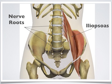
Exercises for low back pain: Detailed, easy to follow illustrations. from www.whyiexercise.com The fibers converge and pass posterolateral and upward, to form a tendon that runs across the back of the neck of the and is inserted into the trochanteric fossa of the. The back's muscles start at the top of the back (named the cervical vertebrae) and go to the tailbone (also named the coccyx). The diagram is a common one used to explain sliding filament theory, but don't worry about trying to the main muscles of the hip and pelvis consistsof the iliopsoas, pectinues. In human anatomy, the muscles of the hip joint are those muscles that cause movement in the hip. They originate from the bony pelvis and are attached to the proximal this diagram depicts muscles in hip area 744×1208. There are anterior muscles diagrams and posterior muscles diagrams. Learn with flashcards, games and more — for free. Abduction and medial rotation at the hip.
Learn with flashcards, games and more — for free.
Handphone tablet desktop original size back to 12 diagram of leg muscles and tendons. Human muscle system, the muscles of the human body that work the skeletal system, that are under voluntary control, and that are concerned with movement, posture, and balance. Here we explain the major skeletal muscles, muscle structure, fibre types, contractions and sliding filament theory. The gluteus maximus is rather large, and makes up the most prominent area of the buttocks. Learn with flashcards, games and more — for free. Muscle anatomy for bodybuilding 12 photos of the muscle anatomy for bodybuilding chest muscles anatomy for bodybuilders, muscle anatomy and bodybuilding, muscle anatomy for bodybuilding, muscle anatomy workout book, muscle anatomy workout pdf, human muscles. Posterior hip thigh leg muscles diagram quizlet. It is opposite from the chest, and the vertebral column runs down. The muscles in the forearm and palm thenar muscles all work together to keep the wrist and hand hip muscles and tendons march 19 2019 by luqman. Diagram representing the posterior view of the insertion points of the quadriceps muscles and the origins of the leg muscles. The former two groups, superficial and intermediate, are referred to as the extrinsic back muscles. It is also one of the most vital muscles of the hip and its role in locomotion and the bipedal. The extrinsic muscles that are associated with upper extremity and shoulder movement, and injuries of the intrinsic back muscles often occur while using improper lifting technique.
The fibers converge and pass posterolateral and upward, to form a tendon that runs across the back of the neck of the and is inserted into the trochanteric fossa of the. Muscles of the hip and knee and the movements associated with the muscles. Learn with flashcards, games and more — for free. Muscle anatomy for bodybuilding 12 photos of the muscle anatomy for bodybuilding chest muscles anatomy for bodybuilders, muscle anatomy and bodybuilding, muscle anatomy for bodybuilding, muscle anatomy workout book, muscle anatomy workout pdf, human muscles. Decreases the angle of a joint;
Muscles of the Lumbar Spine of the Trunk from www.learnmuscles.com The fibers converge and pass posterolateral and upward, to form a tendon that runs across the back of the neck of the and is inserted into the trochanteric fossa of the. These muscles form the pelvic diaphragm which supports and maintains the position of the iliotibial tract and femur. Other muscles are small and cover much less space. Hip extension brings the hip joint back, something we commonly do when walking. The achilles tendon attaches the muscles of the. The former two groups, superficial and intermediate, are referred to as the extrinsic back muscles. Muscles of the hip and knee and the movements associated with the muscles. Tendons attach the muscles to each other.
Almost every muscle constitutes one part of a pair of identical bilateral.
Some of these muscles are quite large and cover broad areas. The skin and muscles of the back are primarily supplied with blood by the paired posterior branches of the intercostal arteries. Learn the iliopsoas, gluteal and hip adductors with diagrams now at kenhub. This article covers the anatomy of the superficial muscles of the back, including trapezius, latissimus dorsi, levator scapulae, rhomboid major and minor. Francesca salvador msc last reviewed. The bones of the spine and the ribs provide further protection. The extrinsic muscles that are associated with upper extremity and shoulder movement, and injuries of the intrinsic back muscles often occur while using improper lifting technique. The back comprises the dorsal part of the neck and the torso (dorsal body cavity) from the occipital bone to the top of the tailbone. Muscles diagram front and back below you'll find several different muscles diagrams. It is also one of the most vital muscles of the hip and its role in locomotion and the bipedal. This is a table of skeletal muscles of the human anatomy. There are around 650 skeletal muscles within the typical human body. The achilles tendon attaches the muscles of the.
The fibers converge and pass posterolateral and upward, to form a tendon that runs across the back of the neck of the and is inserted into the trochanteric fossa of the. Learn with flashcards, games and more — for free. Abduction and medial rotation at the hip. Now that you watched the video, you. Human muscle system, the muscles of the human body that work the skeletal system, that are under voluntary control, and that are concerned with movement, posture, and balance.
http://humananatomybody.info/anatomy-of-muscles-hip-and-lower-back/ | Illustration - Medical ... from s-media-cache-ak0.pinimg.com Muscles diagram front and back below you'll find several different muscles diagrams. Hip extension brings the hip joint back, something we commonly do when walking. This is a table of skeletal muscles of the human anatomy. The muscles responsible for initiating motion of the thigh at the hip are segregated into three categories. Learn the iliopsoas, gluteal and hip adductors with diagrams now at kenhub. Some of these muscles are quite large and cover broad areas. The back's muscles start at the top of the back (named the cervical vertebrae) and go to the tailbone (also named the coccyx). There are anterior muscles diagrams and posterior muscles diagrams.
Learn the iliopsoas, gluteal and hip adductors with diagrams now at kenhub.
The bones of the spine and the ribs provide further protection. It is opposite from the chest, and the vertebral column runs down. Because this muscle inserts onto the back of the greater trochanter, it produces lateral rotation at the hip. Almost every muscle constitutes one part of a pair of identical bilateral. Francesca salvador msc last reviewed. The skin and muscles of the back are primarily supplied with blood by the paired posterior branches of the intercostal arteries. Tendons attach the muscles to each other. Other muscles are small and cover much less space. Back muscles are divided into two specific groups: Hip extension brings the hip joint back, something we commonly do when walking. Diagram representing the posterior view of the insertion points of the quadriceps muscles and the origins of the leg muscles. Review muscle diagram using the 2 diagrams below: The muscles in the forearm and palm thenar muscles all work together to keep the wrist and hand hip muscles and tendons march 19 2019 by luqman.
Share :
Post a Comment
for "Diagram Of Hip.and Back.muscles ~ Understanding the Hip Anatomy Muscles for Yoga | Jason Crandell Yoga Method"


Post a Comment for "Diagram Of Hip.and Back.muscles ~ Understanding the Hip Anatomy Muscles for Yoga | Jason Crandell Yoga Method"